Share on Google+Share on FacebookShare on LinkedInShare on TwitterShare on DiggShare on Stumble Upon
Custom Search


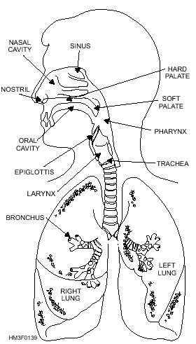
Trachea
The trachea, or windpipe, begins at the lower end of the larynx and terminates by dividing into the right and left bronchi. It is a long tube composed of 16 to 20 C-shaped cartilaginous rings, embedded in a fibrous membrane, that support its walls, preventing their collapse (fig. 1-39).

The trachea has a ciliated mucous membrane lining that entraps dust and foreign material. It also propels secretions and exudates from the lungs to the pharynx, where they can be expectorated.

Bronchi
The bronchi are the terminal branches of the trachea, which carry air to each lung and further divide into the bronchioles.

Bronchioles
The bronchioles are much smaller than the bronchi and lack supporting rings of cartilage. They terminate at the alveoli (fig. 1-40).

Alveoli
The alveoli are thin, microscopic air sacs within the lungs (fig. 1-40). They are in direct contact with the pulmonary capillaries. It is here that fresh oxygen exchanges with carbon dioxide by means of a diffusion process through the alveolar and capillary cell walls

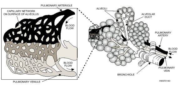
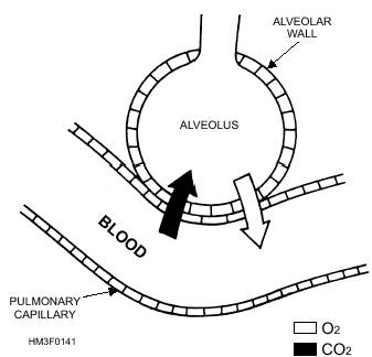
Figure 1-39.-Organs of the respiratory system.

(fig. 1-41). The lungs are cone-shaped organs that lie in the thoracic cavity. Each lung contains thousands of alveoli with their capillaries. The right lung is larger than the left lung and is divided into superior, middle, and inferior lobes. The left lung has two lobes, the superior and the inferior.

Pleurae
The pleurae are airtight membranes that cover the outer surface of the lungs and line the chest wall. They secrete a serous fluid that prevents friction during movements of respiration.

Mediastinum
The mediastinum is the tissue and organs of the thoracic cavity that form a septum between the lungs. It extends from the sternum to the thoracic vertebrae and from the fascia of the neck to the diaphragm. The mediastinum contains the heart, the great blood vessels, the esophagus, a portion of the trachea, and the primary bronchi.

Diaphragm
The diaphragm is the primary muscle of respiration. It is a dome-shaped muscle and separates the thoracic and abdominal cavities. Contraction of this muscle flattens the dome and expands the vertical diameter of the chest cavity.

Intercostal Muscles
The intercostal muscles are situated between the ribs. Their contraction pulls the ribs upward and outward, resulting in an increase in the transverse diameter of the chest (chest expansion).

Inhalation is the direct result of the expansion caused by the action of the diaphragm and intercostal muscles. The increase in chest volume creates a negative (lower than atmospheric) pressure in the pleural cavity and lungs. Air rushes into the lungs through the mouth and nose to equalize the pressure.
Exhalation results when the muscles of respiration relax. Pressure is exerted inwardly as muscles and

Figure 1-40.-Bronchiole and alveoli.

Figure 1-41.-Pulmonary exchange at alveolus. 

bones return to their normal position, forcing air from the lungs.


Western Governors University


Privacy Statement - Copyright Information. - Contact Us

Integrated Publishing, Inc. - A (SDVOSB) Service Disabled Veteran Owned Small Business