|
MANDIBULAR CENTRAL INCISORS The mandibular central incisor (tooth #24 or #25) is illustrated in figure 4-30. These are the first permanent teeth to erupt, replacing deciduous teeth, and are the smallest teeth in either arch. Facial Surfaces\The facial surface of the mandibular central incisor is widest at the incisal edge. Both the mesial and the distal surfaces join the incisal surface at almost a 90 angle. Although these two surfaces are nearly parallel at the incisal edge, they converge toward the cervical margin. The developmental grooves may or may not be present. When present, they appear as very faint furrows.
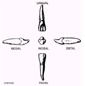
Figure 4-30.\Surfaces of a mandibular central incisor. Lingual Surface\The lingual surface is concave from the incisal edge to the cervical margin. Root Surface\The root is slender and extremely flattened on its mesial and distal surfaces. MANDIBULAR LATERAL INCISORS The mandibular incisor (tooth #23 or #26) illustrated in figure 4-31, is a little wider mesiodistal than the mandibular central incisor, and the crown is slightly longer from the incisal edge to the cervical line. Facial Surface\The facial surface is less symmetrical than the facial surface of the mandibular central incisor. The incisal edge slopes upward toward the mesioincisal angle, which is slightly less than 90. The distoincisal angle is rounded. The mesial border is more nearly straight than the distal border. Lingual Surface\The lingual surface is similar in outline to the facial surface. The incisal portion of the lingual surface is concave. The cingulum is quite large but blends in smoothly with the rest of the surface. Root Surface\The root is single and extremely flattened on its mesial and distal surfaces. MAXILLARY CUSPIDS The maxillary cuspid (tooth #6 or #11) is illustrated in figures 4-32 and 4-33. The maxillary cuspid is usually the longest tooth in either jaw. Since it resembles a dog's tooth, it is sometimes called the canine. Facial Surface\The facial surface of the crown (fig. 4-33) differs considerably from that of the maxillary central or lateral incisors. In that the incisal edges of the central and lateral incisor are nearly
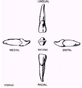
Figure 4-31.\Surfaces of a mandibular lateral incisor.
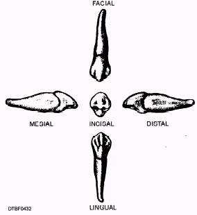
Figure 4-32.\Surfaces of a maxillary cuspid.
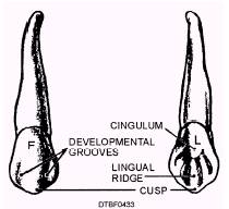
Figure 4-33.\Features of facial and lingual surfaces of a maxillary cuspid. straight, the cuspid has a definite point, or cusp. There are two cutting edges, the mesioincisal and the distoincisal. The distoincisal cutting edge is the longer of the two. The developmental grooves that are so prominent on the facial surface of the central incisor are present here, extending two-thirds of the distance from the tip of the cusp to the cervical line. Lingual Surface\The lingual surface has the same outline as the facial surface but is somewhat smaller because the mesial and distal surfaces of the crown converge toward the lingual surface. The lingual surface is concave, with very prominent mesial and distal marginal ridges, and a lingual ridge, which extends from the tip of the cusp toward the cervical
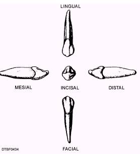
Figure 4-34.\Surfaces of a mandibular cuspid. line. There is often a cingulum in the cervical portion of the lingual surface of the crown. Root Surface\The root is single and is the longest root in the arch. It is usually twice the length of the crown. This is because the cuspid is designed for seizing and holding food.
|

