|
MANDIBULAR SECOND BICUSPID The mandibular second bicuspid (tooth #20 or #29), illustrated in figure 4-39, is the fifth tooth from the midline.

Figure 4-37.\Surfaces of maxillary second bicuspid.

Figure 4-38.\Surfaces of mandibular first bicuspid.

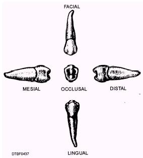
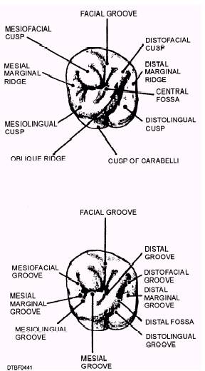
Figure 4-39.\Surfaces of mandibular second bicuspid. Facial Surface\The facial surface has the same facial surface as the first bicuspid. Lingual Surface\The lingual surface is similar to that of the mandibular first bicuspid, with the exception that there may be two lingual cusps present. Occlusal Surface\The occlusal surface usually has a total of three well-defined cusps. Viewed from above, the three cusp present a Y-form pattern. Root Surface\The root of the tooth is single, and in a great many instances, the apical region is found to be quite curved. MAXILLARY FIRST MOLAR The maxillary first molar (tooth #3 or #14), illustrated in figures 4-40 and 4-41, is the sixth tooth from the midline. The first molars are also known as 6-year molars, because they erupt when a child is about 6 years old Facial Surface\The facial surface has a facial groove that continues over from the occlusal surface, and runs down to the middle third of the facial surface. Lingual Surface\In a great many instances, there is a cusp on the lingual surface of the mesiolingual cusp. This is a fifth cusp called the cusp of Carabelli, which is in addition to the four cusps on the occlusal surface. Occlusal Surface\In all molars the patterns of the occlusal surface (fig. 4-41) are quite different from those of the bicuspids. The cusps are large and prominent, and the broad grinding surfaces are broken up into rugged appearing ridges and well-defined grooves. An oblique ridge, which is not present on the bicuspids, appears here (it also appears on maxillary second and third molars). Roots\The maxillary first molar has three roots, which are named according to their locationsmesiofacial, distofacial, and lingual (or palatal root). The lingual root is the largest.

Figure 4-40.\Surfaces of maxillary first molar.

Figure 4-41.\Features of occlusal surfaces of maxillary first molar.
|

