URINARY BLADDER
The urinary bladder functions as a temporary
reservoir for urine. The bladder possesses features that
enable urine to enter, be stored, and later
be released for evacuation from the
body.
Structure
The bladder is a hollow, expandable, muscular
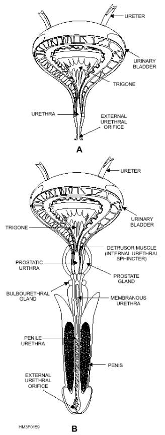
organ located in the pelvic girdle (fig. 1-59). Although
the shape of the bladder is spherical, its
shape is altered by the pressures of
surrounding organs. When it is empty,
the inner walls of the bladder form folds. But as
the bladder fills with urine, the walls become
smoother.
The internal floor of the bladder includes a
triangular area called the trigone (fig. 1-59). The
trigone has three openings at each of its
angles. The ureters are attached to the
two posterior openings. The anterior
opening, at the apex of the trigone, contains a
funnel-like continuation called the neck of the
bladder. The neck leads to the urethra.
The wall of the bladder consists of four bundles of
smooth muscle fibers. These muscle fibers,
interlaced, form the detrusor muscle
(which surrounds the bladder neck)
and comprise what is called the internal
urethral sphincter. The internal urethral sphincter
prevents urine from escaping the bladder
until the pressure inside the bladder
reaches a certain level.
Parasympathetic nerve fibers in the detrusor muscle
function in the micturition (urination)
process. The

Figure 1-59.-Urinary bladder and urethra:
A. Frontal section of the female urinary bladder and urethra;
B. Frontal section of the male urinary bladder and urethra.
outer layer (serous coat) of the bladder wall consists of
two types of tissue, parietal peritoneum and
fibrous connective tissue.
|

